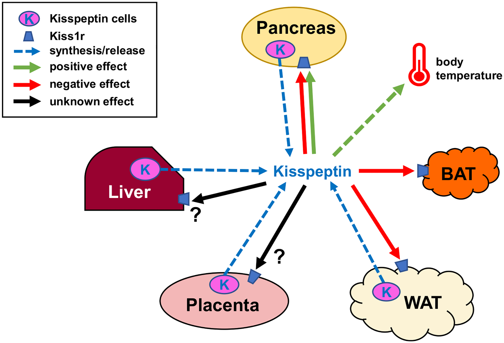
Fig. 5.

Schematic showing possible metabolic actions of peripheral kisspeptin signaling. Blue dotted arrows show potential peripheral tissue sources of circulating kisspeptin the blood. Green arrows denote positive effects of kisspeptin on target tissues or cells, red arrows denote negative effects, while black arrows denote unknown/unstudied effects. Kiss1r is expressed in a wide variety of target tissues including the brain (not shown), liver, pancreas, adipose, BAT, gonad, and placenta (Brown et al., 2008; Hauge-Evans et al., 2006; Herbison et al., 2010; Kotani et al., 2001; Tolson et al., 2020). Kisspeptin signaling has been shown to affect several of these tissues, although for most peripheral effects the endogenous physiological source (specific tissue or cell type) of kisspeptin is unknown. Moreover, it is not clear if there are also autocrine/paracrine effects of kisspeptin acting on the same peripheral tissue that synthesized it. Indeed, the role of kisspeptin synthesis and possible secretion from many peripheral tissues has not yet been studied; for example, the pancreas and white adipose tissue both express Kiss1 and presumably synthesize kisspeptin (Brown et al., 2008; Cockwell et al., 2013; Dudek et al., 2016; Ohtaki et al., 2001; Tolson et al., 2020) but it is presently unclear where such kisspeptin acts or what its specific role(s) might be.
