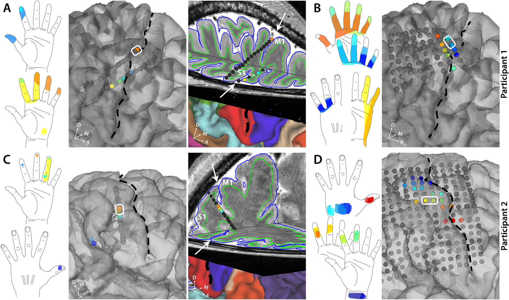
Fig. 2. Self-reported sensory percepts in the hand upon stimulation in S1 sulcal (SEEG) or gyral (HD-ECoG) areas.

A. All the sensory percepts reported by participant 1 upon sulcal stimulation through SEEG electrodes. The color of each electrode matches the color of the corresponding percept evoked. The third panel shows a 3D brain slice showing the same SEEG electrodes. B. All the sensory percepts reported by participant 1 upon gyral stimulation through HD-ECoG electrodes.C.All the sensory percepts reported by participant 2 upon sulcal stimulation through SEEG electrodes. The color of each electrode matches the color of the corresponding percept evoked. The third panel shows a 3D brain slice showing the same SEEG electrodes. Note the more posterior SEEG lead is not shown only in this panel but in Supplementary Fig. S3. D. All the sensory percepts reported by participant 2 upon gyral stimulation through HD-ECoG electrodes. An example bipolar electrode is shown (white rectangle). Black dashed line and white arrows denote the central sulcus.
