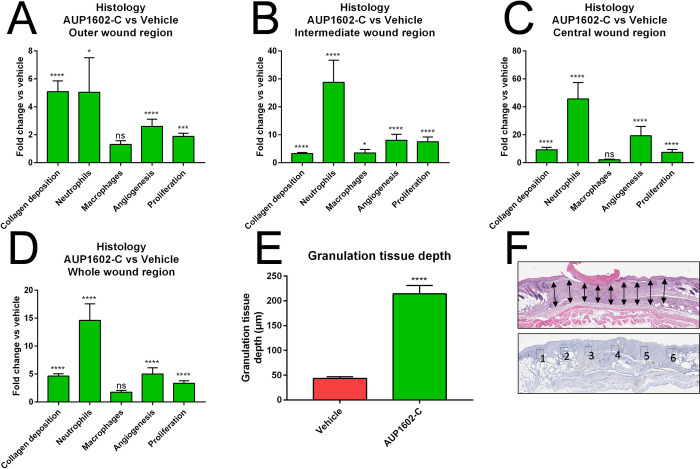
Fig 5. The protein expression fold changes in collagen deposition, neutrophile count, macrophage presence, angiogenesis and cellular proliferation.
Fold changes measured from histological and immuno-histological staining’s after treatment with AUP1602-C in A) outer wound region, B) intermediate wound region, C) central wound region, D) whole wound area. In addition, E) the granulation tissue depth was analysed from the whole wound area. F) The schematic indicating 9 selected locations for measuring the granulation tissue depth as well as definitions of the outer (1 & 6), intermediate (2 & 5), central (3 & 4) and whole (1–6) wound regions. All data are presented as mean ± SEM. Statistical analysis is performed with Kruskall Wallace multivariate analysis followed by ad hoc two sample Mann Whitney U-test analysis). ns = non-significant, *p < 0.05, **p < 0.01, ***p < 0.001 and ****p < 0.0001.

