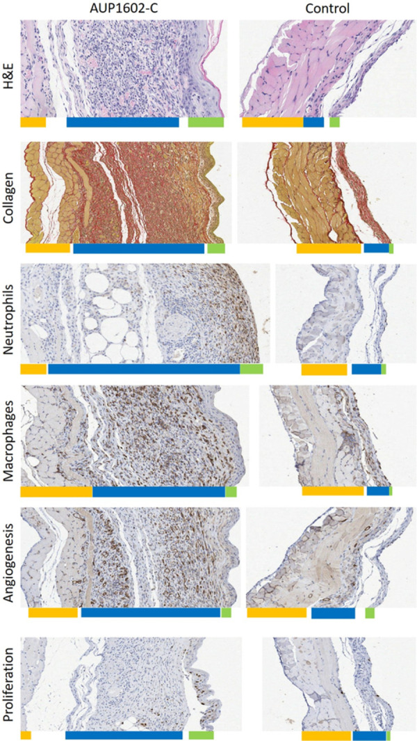
Fig 6. Example microscopic images of each histological and immunohistochemical staining.

Wounds treated with AUP1602-C (left) or vehicle (right) with coloured bars below the sections approximating the thickness of the different tissue layers (yellow: muscle tissue, blue: granulation tissue and green: epithelium. A) H&E staining for GTP depth, B) PSR-staining for collagen deposition, C) anti-CD31-staining for angiogenesis, D) anti-F4/80-staining for macrophages, E) anti-NIMP-R14-staining for neutrophils and F) BrdU staining for cellular proliferation.
