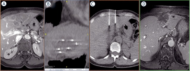
Figure 1.
(A) Solitary liver metastasis from a breast carcinoma in a challenging location between the left and right lobes of the liver, not amenable to surgical resection and progressive under various lines of systemic chemotherapy. The dimensions of the metastasis in segment IVa/b adjacent to segment VIII were 4 x 7 x 5 cm (volume 70 cc). (B) Position of the electrodes in the coronary reconstruction. The aim is to achieve the most uniform coverage of the target lesion by the electrodes. (C) Position of the electrodes in axial cross-sectional imaging. This image shows another essential requirement for the therapeutic success of ECT – the parallelism of the electrodes. (D) The most recent imaging control, complete two years after the ECT procedure, shows complete chemoablation of the entire metastasis, thus formally complete remission of the target lesion without residual or marginal recurrence.

