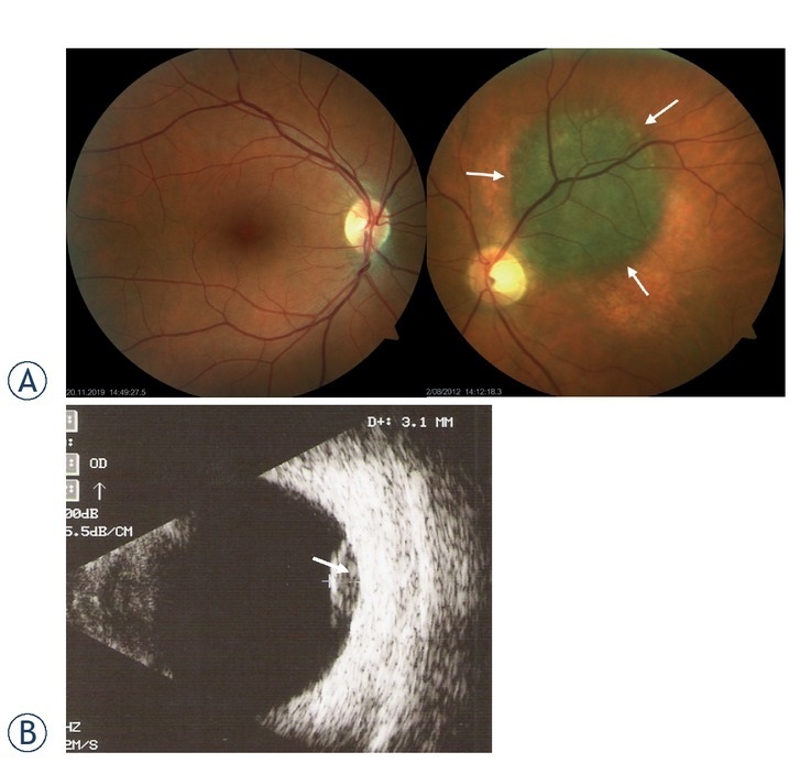
Figure 1.

Ocular fundus of a normal eye, and eye with choroidal melanoma. (A) Ocular fundus. Normal healthy eye (left) versus eye with choroidal melanoma (right; dia = 8 mm and thickness = 2.4 mm). (B) Ultrasound image of the choroidal melanoma (thickness = 3.1 mm).
