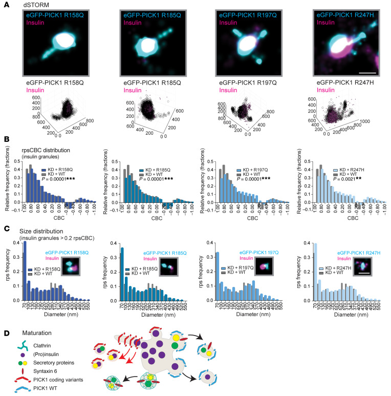
Figure 10. The PICK1 coding variants alter fission from insulin granules.
INS-1E cells were transduced with the coding variants and immunostained for eGFP-PICK (cyan) and insulin (magenta). (A) Representative dSTORM images of eGFP-PICK1 in tubular structures colocalized with insulin. Scale bar: 250 nm. Bottom row: The same data illustrated with 3D; axis values indicate nm. (B) rpsCBC distribution of PICK1 clusters to insulin granules of the 4 coding variants (shades of blue) compared with PICK1 WT (gray). Kolmogorov-Smirnov test was used to test the cumulative distribution, **P < 0.01, ***P < 0.001; n = 4–5 individual experiments. (C) Size distribution of colocalized insulin granules and PICK1 clusters, defined as rpsCBC >0.2, shown with representative dSTORM images of small insulin clusters colocalized with eGFP-PICK1. Scale bar: 250 nm. (D) Proposed model for the PICK1 coding variants in insulin granule biogenesis. We propose that the PICK1 coding variants, with increased abscission efficacy, may cause tubulation and premature budding from the SGs during and/or after the maturation process, generating small clusters that contain excess membrane cargo and insulin.

