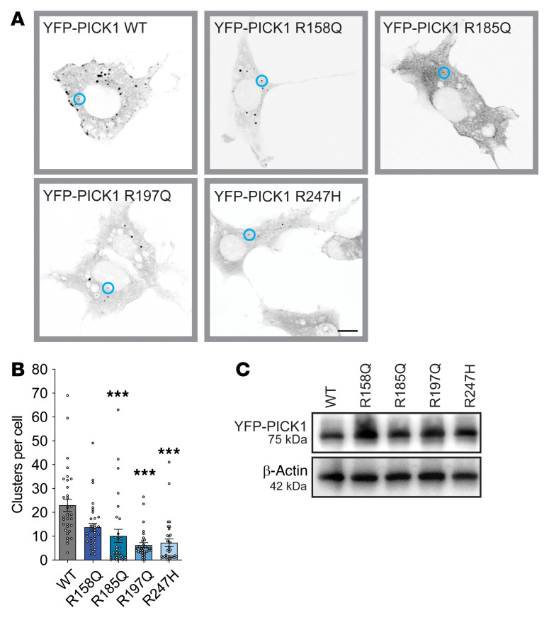
Figure 2. The coding variants display impaired BAR domain function.
(A) COS-7 cells were transiently transfected with YFP-PICK1 WT and the 4 coding variants. Representative confocal images are shown in inverted gray scale; blue circles represent PICK1 clusters. Scale bar: 10 μm. (B) Quantification of clusters per cell. Data are shown as mean ± SEM. Kruskal-Wallis test followed by Dunn’s multiple-comparison test. ***P < 0.001. R158Q (n = 43), R185Q (n = 32), R197Q (n = 31), R247H (n = 40) compared with WT (n = 41). (C) Immunoblotting shows the expression level of YFP-PICK1 WT, R158Q, R185Q, R197Q, and R247H in transiently transfected COS-7 cells.

