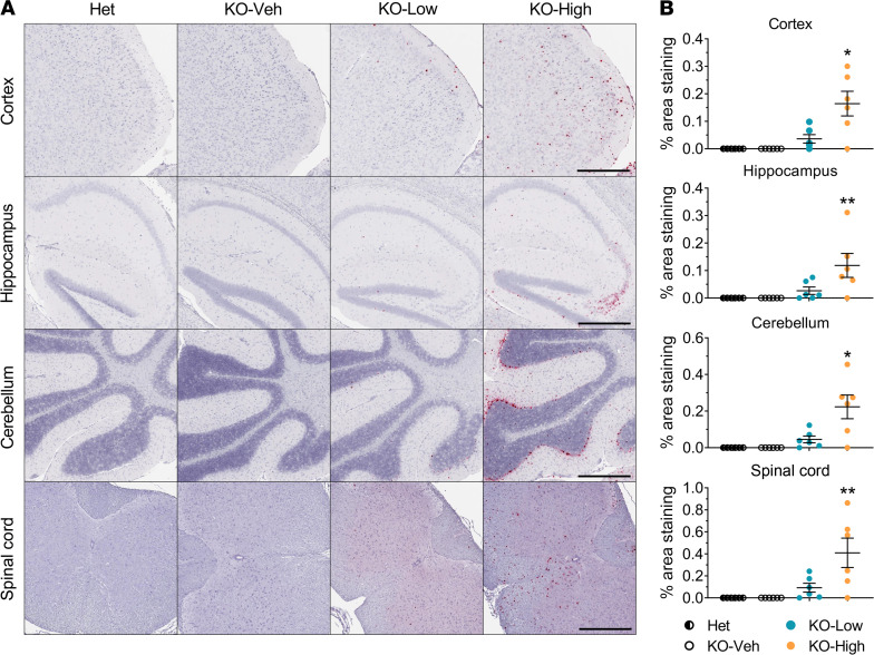
Figure 3. AAV9/MFSD8 GT dose dependently induces hMFSD8opt mRNA expression in the CNS of KO mice.
High (5 × 10¹¹ vg/mouse) or low (1.25 × 10¹¹ vg/mouse) dose of AAV9/MFSD8 vector was administered i.t. to equal numbers of male and female mice at P7–P10. At 4.5 months old, mouse brain and spinal cord were harvested for RNAscope staining to detect hMFSD8opt mRNA (A). Histology images with 1 section/animal were digitized with a ScanScope slide scanner and analyzed using custom analysis settings in HALO Image Analysis Platform. Results are presented as percentage of area staining positive for hMFSD8opt mRNA by tissue region (B). A ROUT test was used first to remove any outlier. Each data point represents a measurement from an individual animal (n = 5–6), with lines representing the mean measurement ± SEM. Data sets that passed tests for normality or homogeneity of variance were analyzed using 1-way ANOVA with α set at 0.05 and Dunnett’s correction for relevant pairwise comparisons. Data sets that did not pass tests for normality or homogeneity of variance were analyzed using the Kruskal-Wallis test with α set at 0.05 and Dunn’s correction for relevant pairwise comparisons. *P < 0.05; **P < 0.01, compared with KO-Veh. Scale bars: 500 μm.

