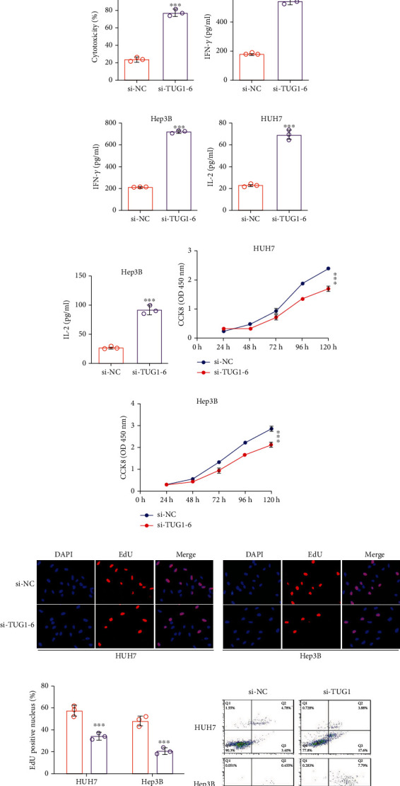
Figure 5.

The antitumor activity of si-TUG1-6 in vitro. (a, b) The immunofluorescence results of HUH7-TUG1-IRES-EGFP and Hep3B-TUG1-IRES-EGFP cells treated with si-TUG1-6. (c) TUG1 expression, (d) Siglec-15 mRNA, and (e, f) protein expression in HUH7 and Hep3B cells treated si-TUG1-6. (g, h) Relative luciferase activity of Jurkat-RGA in HUH7 and Hep3B cells treated with si-TUG1-6. (i, j) T cell-induced cytotoxicity cocultured with HUH7 and Hep3B cells treated with si-TUG1-6. (k, l) IFN-γ and (m, n) IL-2 secreted by T cells from (i, j). (o, p) Cell proliferation in HUH7 and Hep3B cells treated with si-TUG1-6 analyzed by CCK8. (q, r) Cell proliferation in HUH7 and Hep3B cells treated with si-TUG1-6 analyzed by EdU assay. (s, t) Cell apoptosis in HUH7 and Hep3B cells treated with si-TUG1-6 analyzed by Annexin V/PI. (u, v) Apoptosis-relevant proteins level in HUH7 and Hep3B cells treated si-TUG1-6. (w–z) (q, r) Migration and (s, t) invasion of HUH7 and Hep3B cells treated with si-TUG1-6. The data are presented as the means ± SD, n = 3 replicates in (a–v), n = 10 samples in (w–z). ∗P < 0.05, ∗∗P < 0.01, ∗∗∗P < 0.005. S15: Siglec-15.
