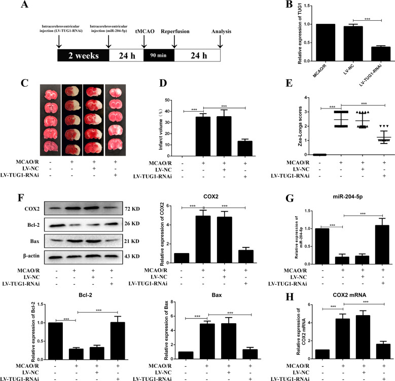
Fig. 2. Knockdown of TUG1 ameliorated brain injury and restrained apoptosis in MCAO/R rats.
A The diagrammatic drawing of in vivo experiment. B The infection efficiency of LV-TUG1-RNAi in cortex of MCAO/R rats (n = 6). C Infarct region was visualized by TTC staining. D Quantitative analysis of brain infarct volume after MCAO/R in rats (n = 9). E Zea-Longa scores (n = 13). F Relative protein levels of COX2, Bcl-2 and Bax in rats (n = 6). Relative expressions of miR-204-5p (G) and COX2 mRNA (H) in rats were detected by qRT-PCR (n = 6). Data are presented as the mean ± SD. ***p < 0.001.

