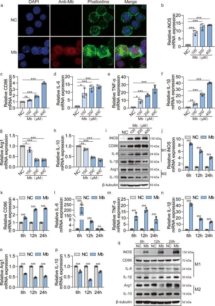
Fig. 2. Myoglobin promotes macrophages polarization to M1 phenotype.
a Representative confocal microscopy images of cells subjected to 200 μM ferrous myoglobin treatments and stained for nuclei (DAPI, blue), anti-myoglobin (red) and microfilament (Phalloidine, green) to detect cytoskeleton (Scale bars: 10 μm). b–i qPCR and WB analyse the expression of M1 molecules iNOS, CD86, IL-6, TNF-α, IL-1β and M2 molecular Arg1, IL-10 in macrophage that treatment with 100, 200, or 400 μM ferrous myoglobin for 6 h. j–q qPCR and WB analyse the expression of M1 molecules iNOS, CD86, IL-6, TNF-α, IL-1β and M2 molecular Arg1, IL-10 in macrophage that treatment with 200 μM ferrous myoglobin for 6 h, 12 h and 24 h, respectively. For statistical analysis, one-way ANOVA followed by Dunnett’s method for multiple comparisons were used in (b–h). Two-way ANOVA followed by Tukey’s method for multiple comparisons used in (j–p). Data are expressed as mean ± SD, n = 3 per group. *P < 0.05, **P < 0.01, ***P < 0.001.

