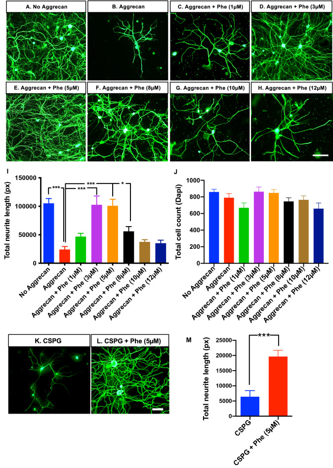
Figure 1.
Effect of phentolamine on DRG neuron outgrowth in vitro. (A–H) Representative images of DRG neurons from 4–6 week old C57BL/6 J mice, cultured for 72 h on the mixture of poly-d-lysine PDL and laminin (Lam) substrates, (A) without aggrecan (B) with aggrecan (C–H) aggrecan with phentolamine (Phe) at concentrations, (C) 1 µM, (D) 3 µM, (E) 5 µM, (F) 8 µM, (G) 10 µM, and (H) 12 µM. Cultured neurons were immunostained with an anti-neuronal marker, β-tubulin III, to identify the neurite outgrowth in DRG neurons (green in color) and imaged with 20 × objective using Olympus microscope (scale bar, 100 µm). (I) Quantification of total neurite length using NeurphologyJ. (J) Quantification of total cell count (based on Dapi staining) using ImageJ. One-way ANOVA with Benjamini and Hochberg false discovery rate correction for multiple comparisons was performed to determine significance among the treatment conditions (n = 4 experiments repeated in duplicates). (K,L) Representative images of DRG neurons (72 h culture), cultured on PDL, and Lam substrates (K) with chondroitin sulfate proteoglycan (CSPG) (L) with CSPG and Phe with 5 µM concentration. (M) Quantification of total neurite length was done as in (I), and an unpaired t-test with Welch’s correction was performed to calculate statistical significance. Data are shown as Mean ± SEM (*p ≤ 0.05, **p ≤ 0.01, ***p ≤ 0.001).

