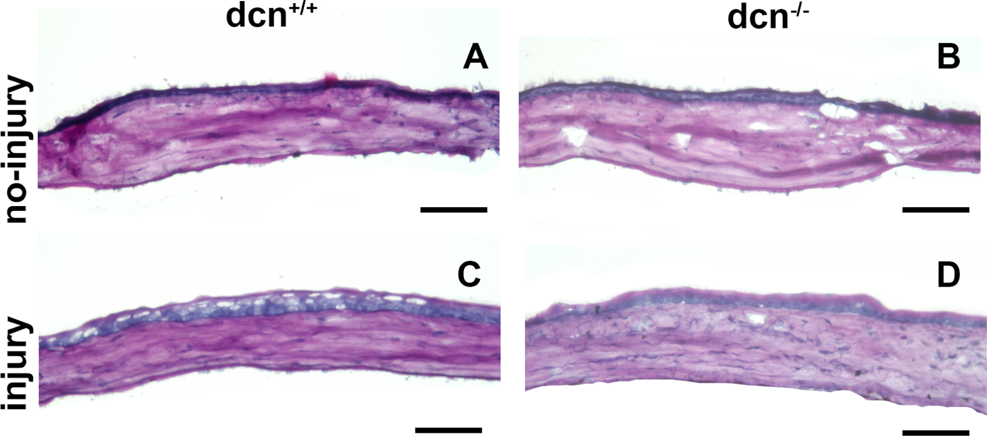
Figure 10.

Representative PAS histology images of no-injury and post-injury dcn+/+ and dcn−/− mouse corneas at day 21. Injured dcn−/− (D) and dcn+/+ corneas (C) showed many morphologic alterations in tissue sections, which were unremarkable in uninjured dcn+/+ (A) and dcn−/− (B) corneas. Scale bar = 100 μm.
