
Figure 2.
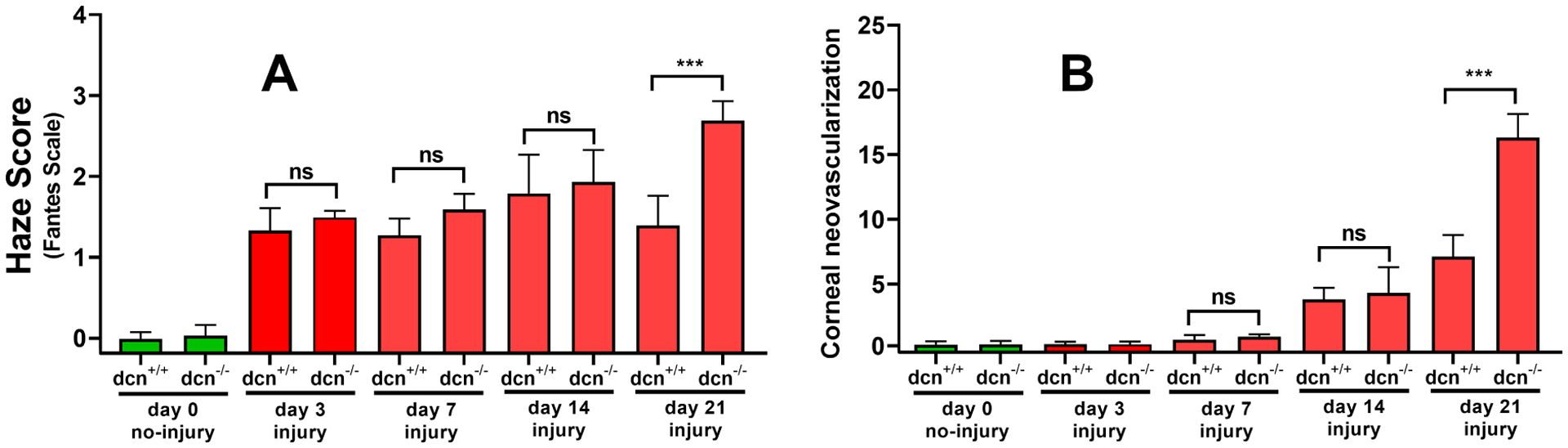
Quantification of corneal haze (A) and neovascularization (B) in uninjured and injured corneas of the dcn+/+ and dcn−/− mice. The corneas of no-injury groups are shown in green bars and injury groups in red bars. Injured dcn−/− corneas showed significantly less haze and neovascularization compared to the dcn+/+ corneas at day 21. Although injured corneas of the dcn+/+ and dcn−/− mice at earlier times (days 3, 7 and 14) exhibited clinically relevant haze and neovascularization but differences between two groups were statistically not significant. Data are expressed in ± SEM and ***P < 0.001.