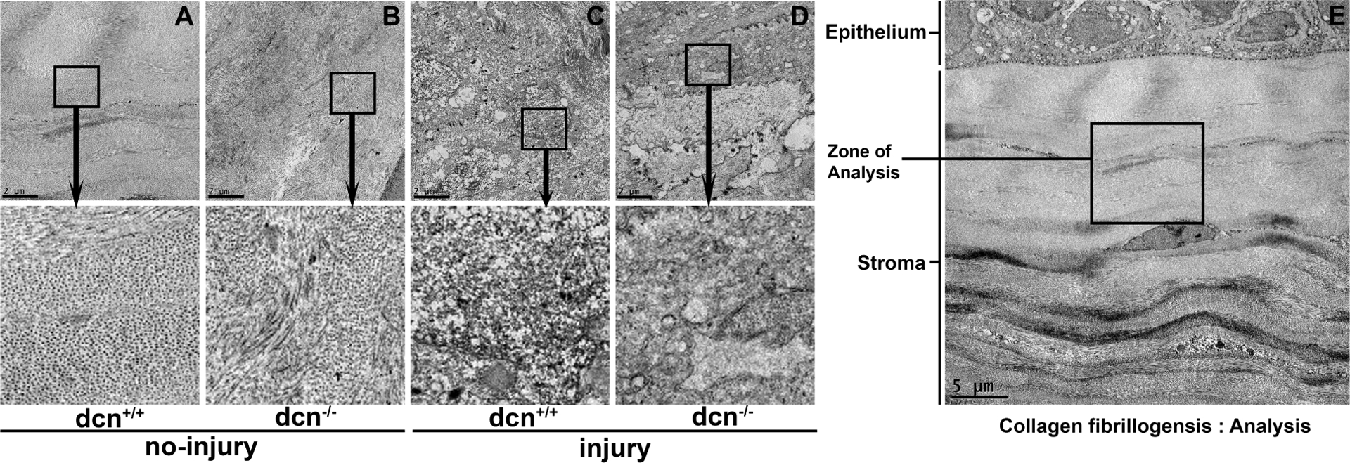
Figure 4.

The TEM images taken from ultrathin sections of no-injury and post-injury corneas of the dcn+/+ and dcn−/− mice at 1500X magnification at day 21. The TEM image (4E; 500X magnification) showing “zone of analysis” in stroma chosen for ultrastructural investigation. Injured dcn+/+ (C) and dcn−/− (D) corneas showed notable disruptions in fibril distribution, organization, and packing in stroma compared to the corresponding no-injury corneas (A & B). The insets show magnified view of selected area. Scale bar = 2 μm (A-D) and 5 μm (E).
