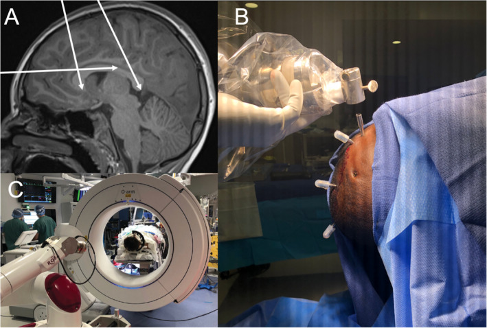
FIGURE 1.

A, Example of a preoperative planning photo using T1‐weighted MRI to visualize LITT catheter trajectory to target the genu, isthmus, and splenium of the corpus callosum. B, Example of capped intraoperative electrode catheters. C, ROSA robot and O‐arm intraoperative imaging system. Not visualized is the Leksell stereotactic system head frame, which is preferred for LITT callosotomy for its flexibility
