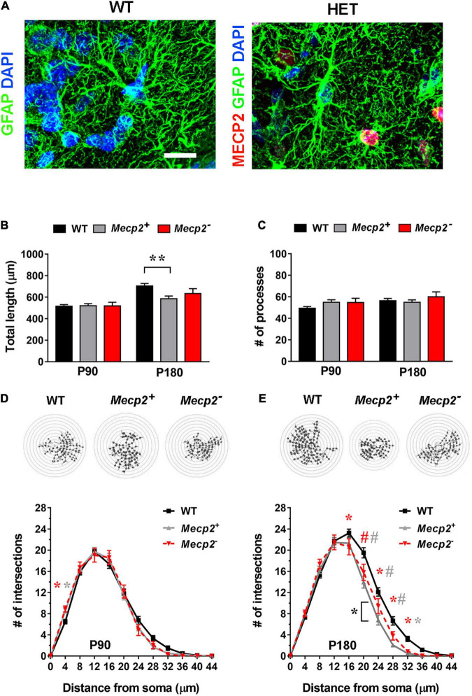
FIGURE 4.
In the layer I of the somatosensory cortex of symptomatic heterozygous animals, the astrocyte cytoskeleton is affected regardless of Mecp2 expression. (A) Micrographs are representative images of astrocytes in the somatosensory cortex (layer I) of WT and heterozygous brains at P180. WT astrocytes were immunostained for GFAP (green) and DAPI (blue); heterozygous brains were also immunostained for Mecp2 (red) to discriminate between cells expressing the WT (Mecp2+) or null (Mecp2–) allele. Scale bar = 10 μm. (B,C) The graphs show the total length (B) and the number of processes (C) in Mecp2+ (gray) and Mecp2– (red) astrocytes derived from the somatosensory cortex (layer I) of P90 and P180 heterozygous female mice, compared to astrocytes of WT animals (black). Data are represented as mean ± SEM. **p < 0.01 by Kruskal–Wallis test, followed by Dunn’s multiple comparison test. (D,E) The graphs depict data from Sholl analysis, reporting the number of intersections of processes of WT (black), Mecp2+ (gray) and Mecp2– (red) astrocytes with concentric circles, at P90 (D) and P180 (E). Representative images of reconstructed astrocyte arbors by SNT plugin are reported above each graph. *p < 0.05, #p < 0.001 by two-way ANOVA, followed by Sidak’s multiple comparison test. * and # indicate the comparison between WT and Mecp2+ astrocytes (gray), WT and Mecp2– astrocytes (red), Mecp2+ and Mecp2– astrocytes (black). Four animals per genotype were used at each time point (N = 4). Astrocytes were randomly selected in the acquired field (P90–110: n = 44 WT, n = 46 Mecp2+, n = 18 Mecp2–; P180: n = 49 WT, n = 35 Mecp2+ and n = 21 Mecp2–; n indicates the number of cells).

