Figure 6.
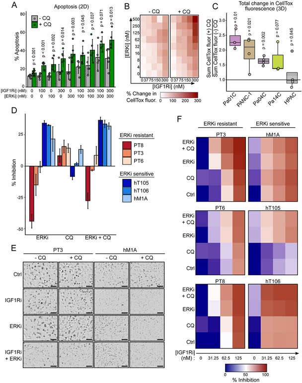
Simultaneous inhibition of ERK, IGF1R and autophagy is cytotoxic in 2D and 3D culture models and inhibits human PDAC organoid growth. A, Flow cytometric determination of apoptosis measured by FITC-annexin V and propidium iodide staining of Pa14C cells treated with CQ (6 μM), BMS-754807 (IGF1Ri), SCH772984 (ERKi), or in multiple combinations (72 hours) as denoted in the figure. Data represent four independent experiments. B, Integrated CellTox fluorescence of Pa14C spheroids was measured following treatment with a matrix of BMS-754807 (IGF1Ri) and SCH772984 (ERKi) in the presence and absence of CQ (6 μM) and CellTox (500 nM, 72 hours). Percent change from DMSO control was calculated for each condition. Data represent average of at least three independent experiments. C, Percent change of CellTox fluorescence was summed across the entire ERKi and IGF1Ri matrix as shown in B for each cell line and normalized by the sum for the (−) CQ control condition. D, Human pancreatic cancer organoids were treated with SCH772984 (500 nM), CQ (10 μM), or the combination (7 days). Data represent the percent inhibition following CellTiter-Glo 3D assay of three independent experiments. E, Representative images of human PT3 and hM1A organoids following treatment with BMS-754807 (IGF1Ri, 31.25 nM), SCH772984 (ERKi, 500 nM), and the combination, with or without CQ (10 μM, 7 days). F, Percent of growth inhibition based on CellTiter-Glo 3D assay of human PDAC organoids treated with BMS-754807 (IGFRi), alone or in combination with CQ (10 μM), SCH772984 (ERKi, 500 nM), or both CQ and SCH772984. Data represent the average of three independent experiments.

