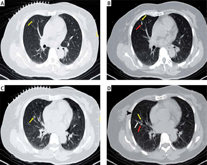
Photo 1.
A 51-year-old woman presented with two lesions in her right lung. The patient underwent simultaneous coil localization for both lesions under CT guidance. A – The CT image shows a lesion in the upper lobe of the right lung (yellow arrow), with a diameter of 8.2 mm and a distance of 6.6 mm from the pleural surface. B – CT image showing a coil (red arrow) adjacent to the lesion in the upper lobe of the right lung (yellow arrow). C – CT image showing a lesion in the middle lobe of the right lung (yellow arrow), with a diameter of 5.0 mm and a distance of 34.5 mm from the pleural surface. D – CT image shows a coil (red arrow) located next to the lesion in the middle lobe of the right lung (yellow arrow), with a small amount of pneumothorax (black arrowhead) and pulmonary hemorrhage (white arrow)

