
Figure 3. GABARρ2 receptors are present at the contact points between horizontal cell bulbs and CBC dendrites.
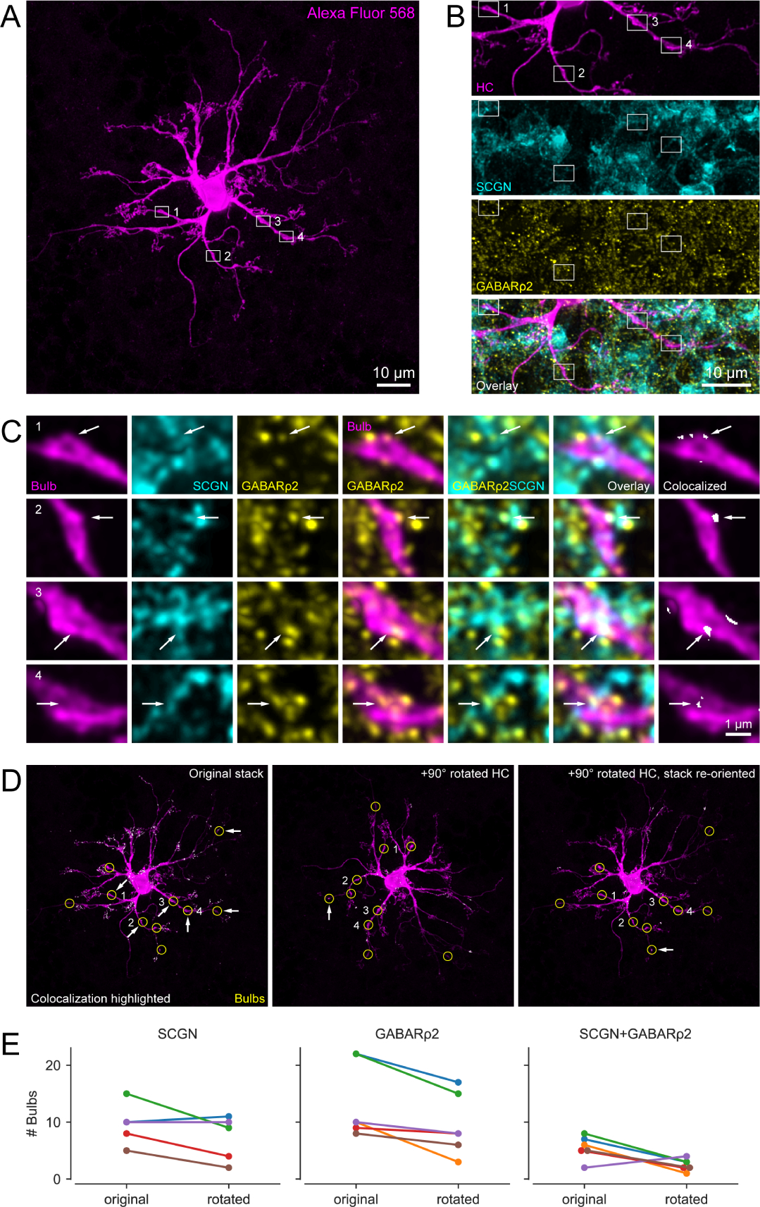
(A) Alexa Fluor 568-injected HC with identified bulbs (white boxes indicate examples). Note that the brightness was increased for better visibility. (B) Magnified clipping from (A) showing four representative bulbs (magenta) as well as their corresponding secretagogin (SCGN, cyan) and GABARρ2 (yellow) stainings. The bottom panel represents overlay of the three channels. (C) Enlarged bulbs from boxes in (B) with SCGN and GABARρ2 immunolabeling. Both individual (columns 1–3) and merged channels (columns 4–6) are shown, with arrows indicating colocalization. Colocalization of all three channels at the bulbs (column 7) identified by ‘colocalization highlighter’ plugin in Fiji (see panel D). (D) Colocalizing points highlighted in original (left) and 90-degree rotated HC (center, right), with bulbs encircled (yellow). Arrows denote bulbs with colocalization. (E) Line plots showing number of bulbs (per HC, n = 6 HCs) colocalizing with SCGN (left), GABARρ2 (center) and both (right) for non-rotated (original) and 90-degree rotated condition, see also Table S1. Note that images are maximum projections: (A, D) of the entire HC stack (85 optical slices), (B) of the selected clipping (40 optical slices), and (C) of 5–7 optical slices for better visibility of the bulbs. Optical section thickness was 0.17μm. Colocalization analysis itself were carried out within each optical section.