
Figure 4. Mitochondrial structure in HC bulbs.
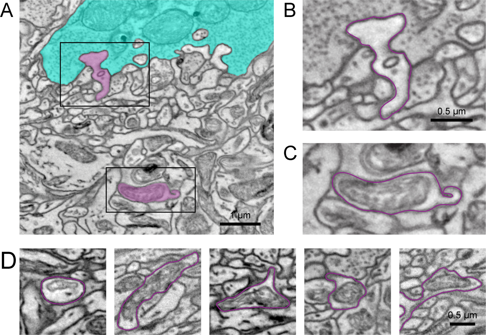
(A) Electron micrograph showing a manually traced HC (magenta) with a dendritic tip invaginating in a cone axon terminal (cyan) (B) and the primary dendrite below the cone axon terminal with a HC bulb of the same cell (C). See also the Video S1 visualizing the complete EM stack. (D) Additional examples of HC bulbs (magenta outline). Note the mitochondrial structure in the bulbs. Black rectangles: location of magnifications shown in (B,C).