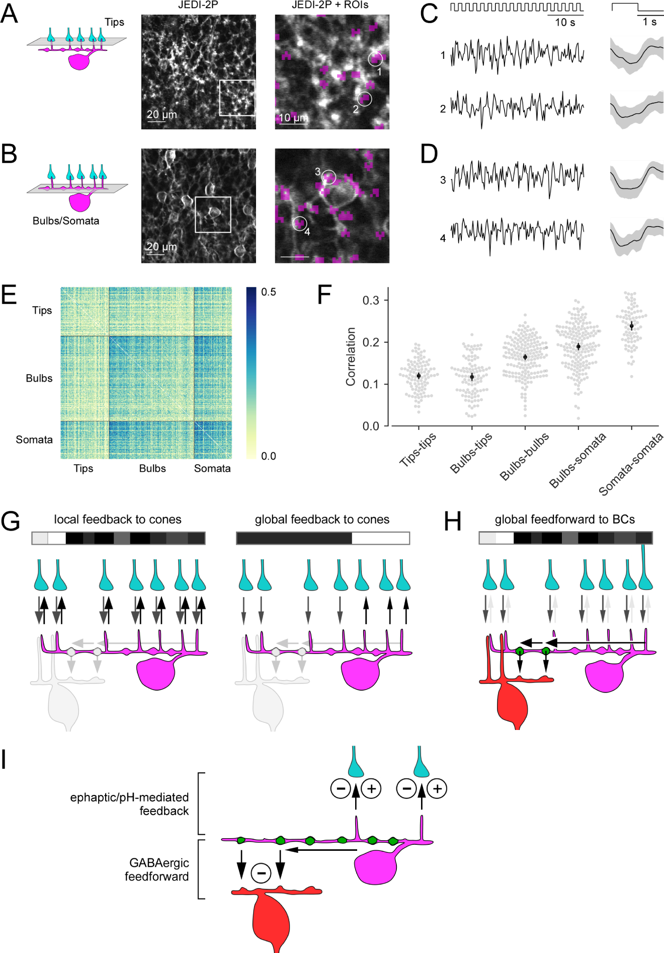
Figure 7. Voltage imaging in the OPL and bilayered synaptic circuitry of horizontal cells in the outer mouse retina.

(A, B) Horizontal (x-y) scans of a Cx57cre/+ transgenic mouse retina in which HCs express the fluorescent voltage sensor JEDI-2P after intravitreal AAV injection (see STAR Methods), with the focal plane in the OPL at the level of the HC tips (A) and of the primary dendrites (B). Right: zoomed-in images of the boxed areas. Regions-of-interest (ROIs, purple) were defined by correlation between neighboring pixels, with ROI size clamped to max. 3 μm in diameter. At the primary dendrite level, ROIs were manually sorted into somata (e.g., ROI 3) and bulbs (e.g. ROIs 1, 2, 4). (C, D) Voltage responses (left, single trials; right, averages with s.d. shading) of representative ROIs to full-field white flashes, with the focal plane in the OPL at the level of the HC tips (C) and at the level of the primary dendrites (D). (E) Correlations between single trials of tips (n = 104), bulbs (n = 181) and somata (n = 80). (F) Mean correlations within and between ROI types. Each dot in the swarm plots (gray) corresponds to one ROI and shows its mean correlation with all ROIs of the reference ROI type (mentioned first in the x-axis labels). Black: Mean +-95%-confidence interval. Except for tips-tips vs. tips-bulbs, all neighboring groups of correlations are significantly different (permutation test, all significant p’s < 0.001, see STAR Methods). (G) Dendritic tips of a HC (magenta) receive cone input (grey arrows) and provide local, cone-specific feedback (black arrows) to cone axon terminals (cyan) in the presence of a spatio-temporally uncorrelated white noise stimulus (white/grey/black bar) (left). For a more spatially correlated stimulus patterns, the feedback to cones may contain a global component (right; local feedback component not shown). (H) For an uncorrelated stimulus (like in G, left), the cone input signals (grey arrows) are integrated in the HC dendrite (black arrow) and forwarded as a global signal by a BC-contacting bulb (green) to the BC (red) forming their surround signal. (I) Illustration of the two synaptic computations performed by HCs at the two distinct OPL strata: Ephaptic/pH-mediated negative and positive feedback (minus/plus symbols) to cone axon terminals in the outer OPL and inhibitory GABAergic feedforward synapses (minus symbol) from HC bulbs to BCs in the inner OPL. GABAergic auto-synapses at the distal dendritic tips of HCs 11 are not shown. Black arrows indicate HC synaptic output.
