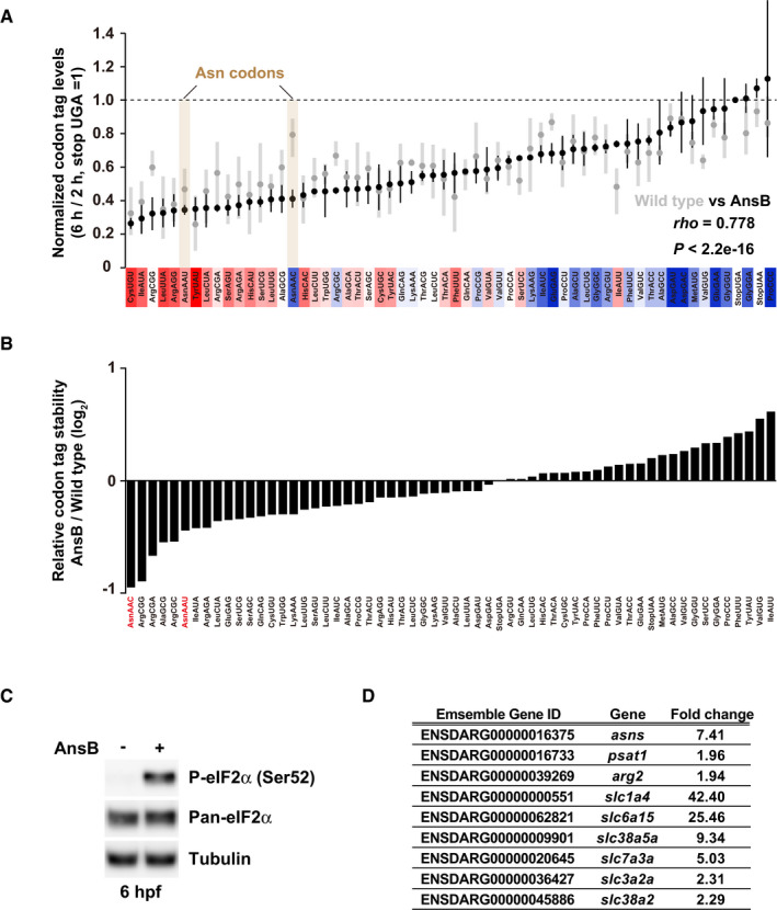
Figure EV3. PACE under asparagine deprivation conditions.

- Results of PACE in AnsB‐expressing zebrafish embryos. Black circles show the relative stability of the reporter mRNAs (averages of two biological replicates) in AnsB‐expressing embryos. Gray circles show the relative stability of reporter mRNAs in wild‐type embryos presented in Fig 2B. The stability of a codon‐tag reporter with a UGA stop codon is set to one. Error bars represent maximum and minimum data points. The relative effect of each codon on mRNA stability in wild‐type embryos measured in Fig 2B is shown as a color gradation with red (destabilizing) to blue (stabilizing). rho, Spearman's correlation. Significance was calculated by Student’s t‐test.
- Relative change in codon‐tag reporter stability between wild‐type and AnsB‐expressing embryos. PACE results in AnsB‐expressing embryos were divided by PACE results in wild‐type embryos and are shown as bar charts. Asn codons are indicated in red.
- Western blotting to detect phospho‐eIF2α (Ser51) and pan‐eIF2α at 6 hpf in the absence or in the presence of AnsB. Tubulin was detected as a loading control.
- A list of genes related to amino acid metabolism or transport and upregulated in AnsB‐expressing embryos compared to wild‐type embryos at 6 hpf. Fold changes assessed by RNA‐Seq and ensemble gene ID are shown.
Source data are available online for this figure.
