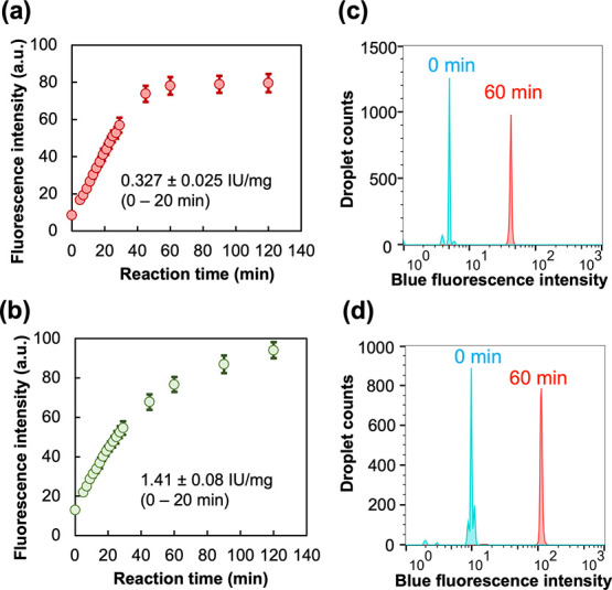
Figure 2.

Detection of purified DPP activity in WODLs using dipeptidyl ACA substrates. (a,c) PmDAPBII-hydrolyzed Met–Leu–ACA. (b,d) PgDPP11-hydrolyzed Leu–Asp–ACA. (a,b) Enzyme reaction curves. Fluorescence intensity was measured from microscopy images with ImageJ.30 Micrographs used in the analysis are shown in Figure S9. Standard deviations were obtained from the fluorescence intensity of 10 droplet images. (c,d) FADS histogram. “0 min” means analyzing immediately after generating the WODLs. A total of about 10,000 droplets were analyzed by FADS.
