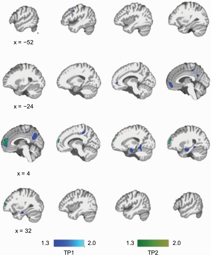
Fig. 1.
Cross-sectional whole-brain voxel-wise GMV comparisons between PMD and NPMD at two time points. PMD showed lower GMV in the left middle frontal gyrus, medial prefrontal cortex (MPFC), praecuneus, right amygdala/hippocampus, and right lingual gyrus at TP1 (before ECT). PMD showed lower GMV in the MPFC at TP2 (after ECT). There were no brain regions that were larger in PMD than NPMD. Significance threshold was set at family-wise error-corrected P < .05 determined by threshold-free cluster enhancement. The color bars represent –log(P) (ie, 1.3 is equivalent to “P = .05”). Abbreviations: ECT, electroconvulsive therapy; GMV, gray matter volume; NPMD, nonpsychotic major depression; PMD, psychotic major depression.

