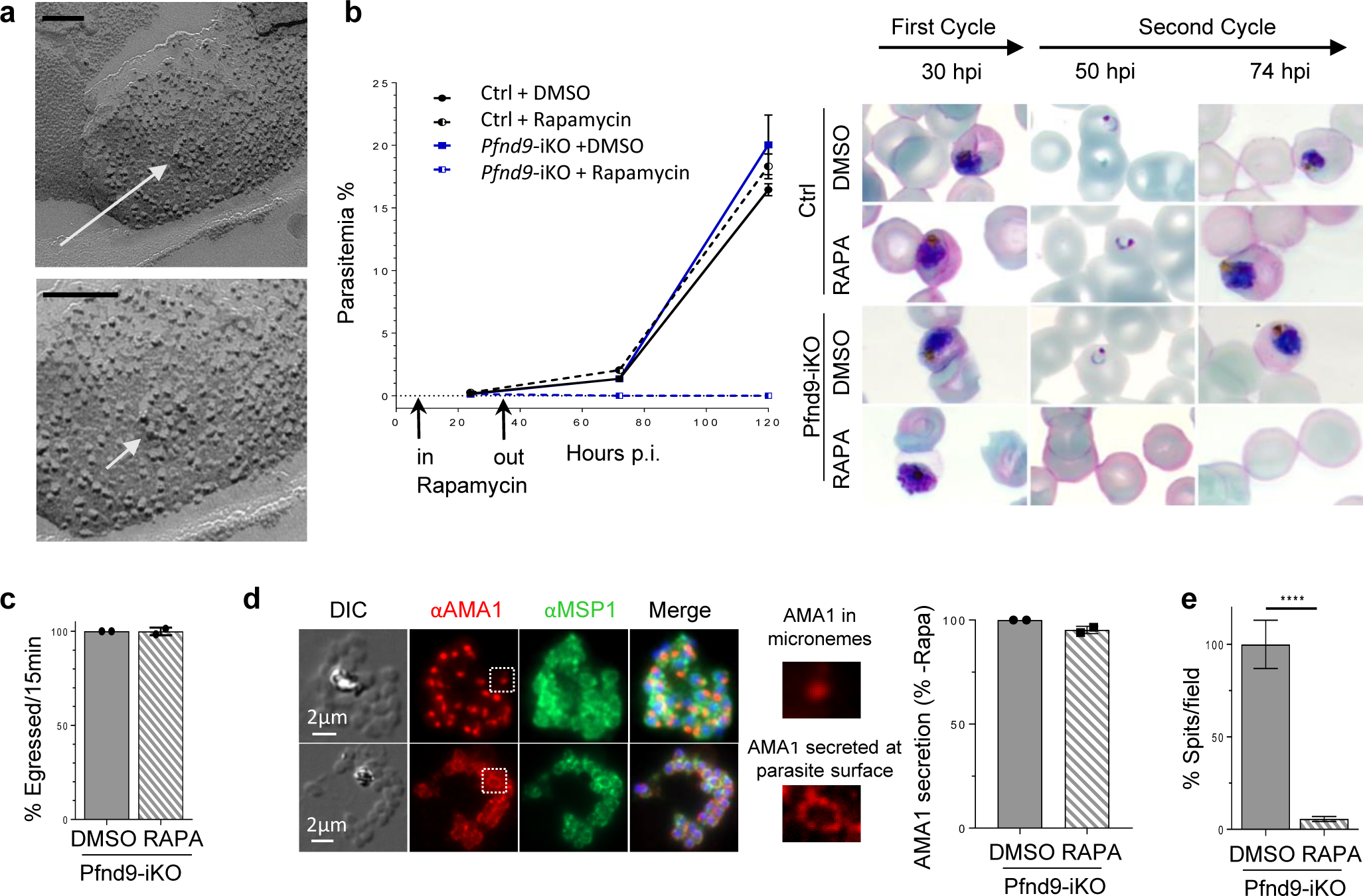
Fig. 2 |. PfNd9 is essential for rhoptry secretion in P. falciparum.

a, Freeze-fracture electron microscopy of a P. falciparum merozoite (P face) showing a rosette of intramembranous particles (white arrow). Higher magnification at the bottom. Bar is 100 nm. b, Growth curves (parasitaemias) of p230p DiCre (Ctrl) and Pfnd9-iKO mutant ± rapamycin shows that PfNd9-depleted parasites have a growth defect. On the right: Giemsa staining of the growth experiment illustrating development and reinvasion of p230p DiCre (Ctrl) and Pfnd9-iKO merozoites (along 2 cycles) ± rapamycin treatment. c, Quantification of egress of Pfnd9iKO ± rapamycin schizonts. Data collected from 8 movies of Pfnd9-iKO ± rapamycin. d, Left: IFA illustrating AMA1 protein stored in micronemes (top) or secreted and translocated at the surface of the parasite prior to egress (bottom). MSP1: surface marker. Right: Proportion of infected cells (± rapamycin) exhibiting AMA1 secretion.. e, Quantification of rhoptry secretion events in Pfnd9-iKO ± rapamycin-treated schizonts using anti-PfRAP2 antibodies to visualise rhoptry secretion events (‘spits’ of RAP2 export into the RBC).
