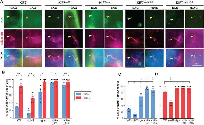
FIGURE 3:
KIF7’s microtubule binding is dispensable for its Hh-induced accumulation at the tip of the cilium. (A) Representative images of the subcellular region containing the primary cilium in Kif7−/− MEFs expressing mCit-tagged KIF7 WT or variant proteins (green) and either untreated (−SAG) or treated with SAG for 4 h (+SAG). The cells were fixed and stained with antibodies against acetylated tubulin (cilium; red), pericentrin (basal body; magenta), and with DAPI (nucleus; blue). Arrowheads indicate tips of cilia. Scale bar, 5 µm. (B–D) Quantification of the percent of cells exhibiting ciliary tip localization of KIF7 WT or variant proteins. The data are plotted to display statistical comparisons (B) between −SAG and +SAG conditions for each expressed protein (two-tailed t test) or (C, D) for the −SAG or +SAG conditions across KIF7 WT and variants (one-way ANOVA with Dunnet’s post hoc test). n.s., not significant; *, p < 0.05; **, p < 0.01; ***, p < 0.001. Each spot indicates the mean of one independent experiment. Error bars, SEM across more than 30 cilia and more than or equal to three independent experiments.

