Figure 1.
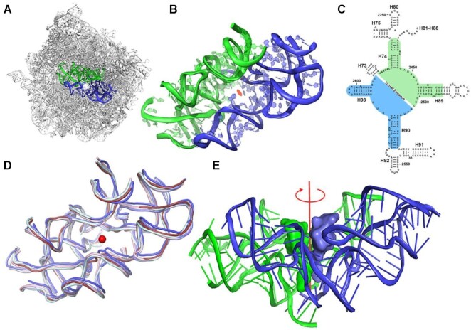
The protoribosome concept. (A) The symmetrical region, marked in blue (A-reg) and green (P-reg), within the rRNA scaffold of the large ribosomal subunit of D. radiodurans (PDBID 1NKW). (B) A close-up of the protoribosome where the 2-fold semi-symmetrical parts are shown. The view is along the pseudo-symmetry 2-fold axis. The center of the PTC is marked by an orange ellipse. (C) A two-dimensional structure diagram of the rRNA surrounding the PTC depicting the symmetry. The A- and P-reg nucleotides are marked using blue and green backgrounds, respectively. 23S rRNA helices numbers are marked in black labels. Nucleotides numbering according to E. coli is shown. (D) Overlay of the symmetrical region of ribosome structures from various organisms representative of various phylogenetic classes: bacterial (D. radiodurans and E. coli in slate and light blue, respectively), Yeast (S. cerevisiae in pale cyan), parasite (L. donovani in blue) and Human ribosomes (cytosolic and mitochondrial in ruby and light pink, respectively) (PDBID used are: 1NKW, 4V4Q, 4V7R, 3JCS, 4U60 and 3J7Y, respectively). The central red dot represents the position of the putative symmetry axis, which is perpendicular to the plane. (E) CCA-3’ end of A-site and P-site tRNAs were superimposed on the symmetrical region of the bacterial ribosome (PDBID 1NKW). The view is perpendicular to the semi-symmetrical 2-fold axis, shown in red.

