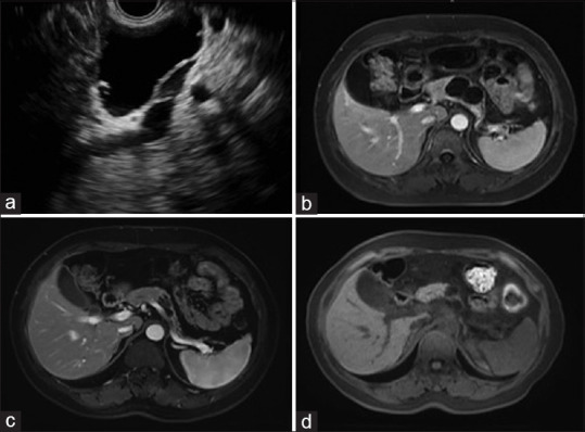
Figure 3.

(a) EUS image before ablation, showing a cystic lesion in the body; (b) magnetic resonance imaging of the same cyst before ablation, showing a 41.0 mm × 27.0 mm cyst; (c) follow-up magnetic resonance imaging at 5 months after the first ablation, showing partial resolution with the cyst decreased to 24.0 mm × 11.0 mm. Then, a second ablation was performed; (d) follow-up magnetic resonance imaging at 17 months after the first ablation, showing that the cyst decreased to 15.0 mm × 12.0 mm
