
Figure 5.
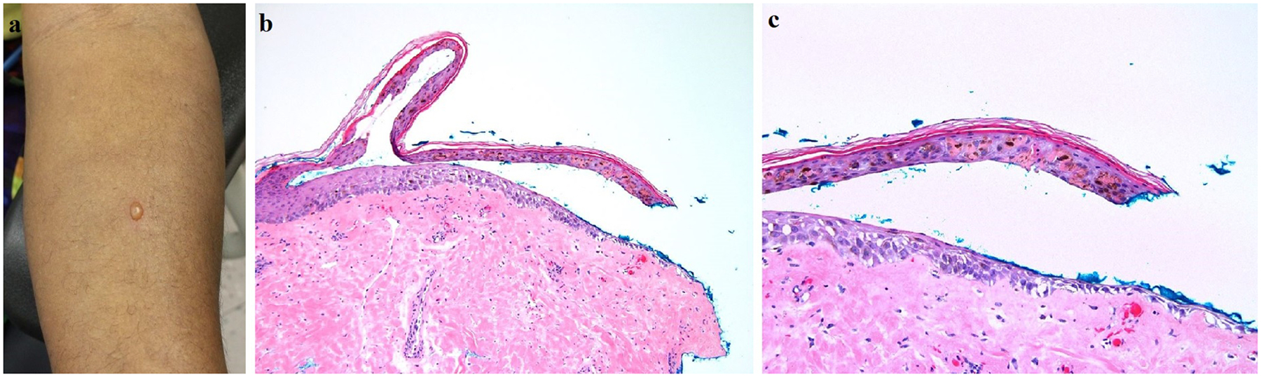
(A) Development of a 5 mm blister on the volar forearm of an African American subject after receiving the first HF-LED-RL treatment session (480 J/cm2). (B) Biopsy of the blister edge shows a subepidermal split with partial re-epithelialization and no significant underlying inflammatory infiltrate (hematoxylin & eosin, 100x). (C) On higher magnification, necrotic keratinocytes within the blister roof are evident, along with clumping of melanin pigment (hematoxylin & eosin, 200x).