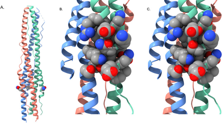
Figure 7.
A) Post-fusion six-helix bundle (PDB ID: 6LXT) illustrating the location of the N1192S mutation. S1192 is rendered as a CPK residue (one per receptor monomer) and color-coded by atom type as described in Figure 5. B) Detailed view of side chain packing interactions formed by N1192. K933 is from the neighboring monomer; this image illustrates the tight packing contacts formed by proximate side chains that stabilize the six-helix bundle fusion structure. C) Detailed view of side chain packing interactions formed by the S1192 mutant. The helix bundle orientation is identical to that in Figure 7B.

