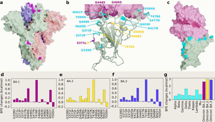
Figure 1:
3D structures of Omicron strains, their ACE2 complexes and their mutation-induced BFE changes. a Spike protein (PDB: 7WK2 [3]) with Omicron mutations being marked yellow. b BA.1 and BA.2 RBD mutations at the RBD-ACE interface (PDB: 7T9L [21]). The shared 12 mutations are labeled in cyan, BA.1 mutations are marked with magenta, and distinct BA.2 mutations are plotted in yellow. b The structure of the RBD-ACE2 complex with mutations on cyan spots. e, f and g BFE changes induced by mutations of Omicron BA.1, BA.2, BA.3, respectively. h a comparison of predicted mutation-induced BFE changes for few SARS-CoV-2 variants.

