Figure 4.

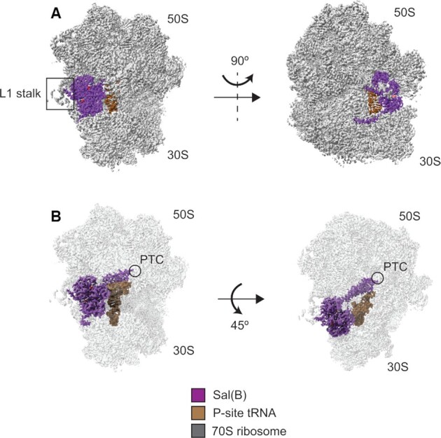
Structure of the Sal(B)•ribosome complex. Sal(B) (purple) binds to the E-site of the ribosome (grey), with the interdomain linker of the former contacting and distorting the P-site tRNA (brown). ATP molecules coloured by atom. (A) Left; front view showing the NBDs of Sal(B) bound to the E site, in close proximity to the L1 stalk. Right; side view showing the C-terminal tail of Sal(B) wrapping around the 30S subunit. (B) Left; front view with transparent ribosome density to show the interdomain linker of Sal(B) reaching towards the peptidyl transferase centre (PTC) of the 50S ribosomal subunit, where LSAP antibiotics bind. Right; top view showing the interaction of the interdomain linker of Sal(B) with the P-site tRNA.
