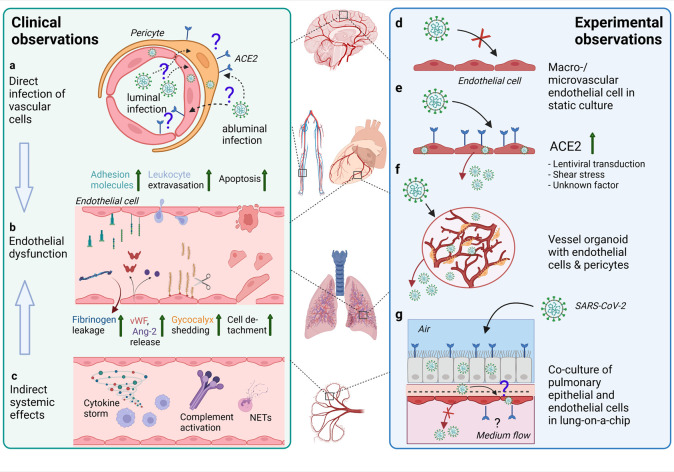
FIGURE 3.
Effects of SARS-CoV-2 infection on the endothelium as observed in patients and different experimental models. (A) In endothelial cells in various organs (such as depicted in the middle of the figure) from COVID-19 patients, SARS-CoV-2 RNA or protein has been inconsistently detected. The ACE2 expression is rather found in pericytes than endothelial cells. Infection of vascular cells can happen from the abluminal side at primary sites of infection in the respiratory tract or from the luminal side in case of hematogenous SARS-CoV-2 dissemination to distal organs. To what extent infection of endothelial cells and/or pericytes occurs and contributes to COVID-19 pathology is unclear. (B) Endothelial dysfunction is not only observed in the pulmonary vasculature, but throughout the body as a hallmark of severe COVID-19 and may be one of the main contributors to increased frequency of thrombotic events. Elevated levels of markers of endothelial activation and injury including von Willebrand factor (vWF), angiopoietin-2 (Ang-2), soluble forms of several cell adhesion molecules and glycocalyx degradation products, and increased numbers of detached endothelial cells are found in COVID-19 patients’ blood. Postmortem analysis of multiple organs has revealed perivascular immune cell infiltrates, decreased endothelial barrier properties as visualized by fibrinogen leakage and endothelial apoptosis in some of the COVID-19 patients. (C) Besides direct infection of vascular cells, immune-mediated mechanisms such as excessive pro-inflammatory cytokine production or complement-hyperactivation can play a major role in causing endothelial dysfunction. (D) Macro- and microvascular endothelial cells isolated from different anatomical locations (such as depicted in the middle of the figure) in conventional monolayer culture were not permissive to infection by SARS-CoV-2. (E) When ACE2 was introduced into endothelial cells by lentiviral transduction productive infection occurred. ACE2 expression in endothelial cells might also be induced by shear stress or certain interferons. (F) In co-culture models such as hiPSC-derived vessel organoids consisting of endothelial cells and pericytes, productive infection occurred, (G) whereas in lung-on-a-chip-models infection of the lung epithelial compartment can lead to infection and cytopathogenic effects of the adjacent endothelial cells (This figure was made with biorender.com).

