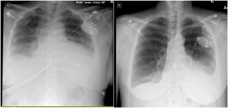
Figure 4. Chest X-rays before and after corticosteroid therapy .
A: Chest X-ray showing left-sided pleural effusion and/or atelectasis/infiltrate, right-sided pleural effusion, and right basilar atelectasis.
B: Chest X-ray after steroids therapy showing decreased left-sided pleural effusion with improved aeration at the left lung base with mild left basal compressive atelectasis and residual effusion, and mild cardiomegaly.

