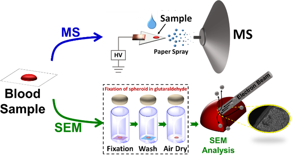
Figure 2.

Analytical experimental workflow involving direct analysis of dry blood by PS MS (top sequence) and scanning electron microscopy analysis following sample fixation with glutaraldehyde (bottom sequence). Blood samples were fixed at different stages of the spheroid drying process to create various snapshots of the condition of red blood cells.
