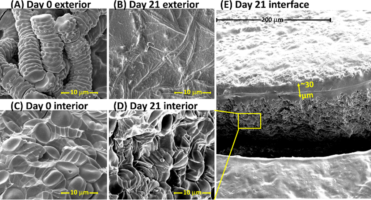
Figure 3.

Scanning electron micrograph of the exterior (A, B) and interior (C, D) of a 10 µL dried blood spheroid, with a micrograph of a large interfacial area shown in E. (A) Exterior surface of a 10 µL dried blood spheroid after drying under ambient storage conditions for 1 h. The rouleau conformation of stacked red blood cells are observed at the spheroids surface after drying the blood for 1 h. (B) Exterior surface of a 10 µL dried blood spheroid after drying under ambient storage conditions for 21 days. No distinct red blood cells are detected. (C) Interior surface of a 10 µL dried blood spheroid after drying under ambient storage conditions for 1 h. Red blood cells persist in clusters and stacks of rouleau; distinct red blood cells are observable. (D) Interior of 10 µL dried blood spheroid after drying under ambient storage conditions for 21 days, zoomed-in from the interfacial photo in E. Red blood cells are still distinguishable and exist in clusters, including stacks of rouleau. (E) Interface showing exterior and interior of a 21-day-old dried blood spheroid. A distinct thin barrier approximately 30 µm of lysed red blood cells is observed that is believed to protect the interior of the dried spheroid.
