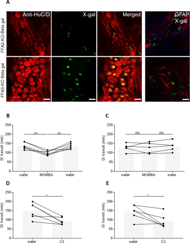
Figure 2. Both FFA2 and FFA3 are expressed in myenteric neurons and promote increased gut transit.
(A) Myenteric plexus dissected from mice expressing a β-galactosidase reporter gene, driven by the Ffar2 (upper panel) or Ffar3 (lower panel) gene promoter sequences were immunostained with anti-HuC/D to identify enteric neurons (left-hand panels, red) and with X-gal to identify receptor-expressing cells (second panels, green). Merged images (third panels) showed coexpression. Myenteric plexus immunostained with X-gal and Glial Fibrillary Acidic Protein (GFAP) to identify enteric glia did not show any coexpression (right-hand panels). Blue: staining with 4′,6-diamidino-2-phenylindole (DAPI) to identify cell nuclei. Scale bar = 20 µm. Male hFFA2-DREADD-HA (B), CRE-MINUS (C), FFA2-KO-βGAL (D), or FFA3-KO-βGAL (E) mice were acclimatized for 7 days with free access to drinking water. Individual animals were then gavaged with carmine red and total GI transit time measured. Following the initial transit studies, mice were provided with 4-methoxy-3-methyl-benzoic acid (MOMBA) (15 mM) or C3 (150 mM) in the drinking water as indicated. After a further 7 days GI transit of all mice was again measured. MOMBA was then removed and the mice were again provided with water followed by a further gavage with carmine red 7 days later. Data are for individual animals (*p < 0.05, **p < 0.01, ns = not significant). One-way analysis of variance followed by Bonferroni’s Multiple Comparison Test.


