
FIGURE 5.
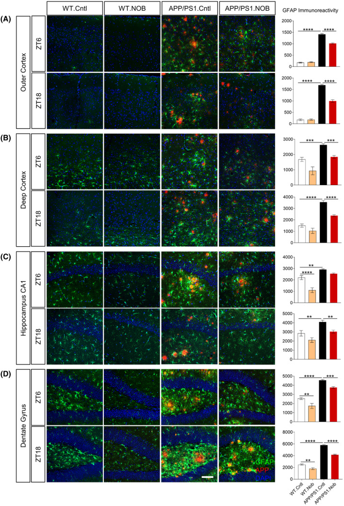
NOB significantly reduces reactive astrocytes in APP/PS1 hippocampus. Double immunofluorescence of astrocytes (GFAP, green) and Aβ (4G8, red) in APP/PS1 mice with DAPI (blue) in the (A) outer cortex, (B) deep cortex, (C) hippocampus CA1, (D) dentate gyrus at two different time points (ZT6 and ZT18). Scale bar: 100 µm. Right Panels: Quantification of the GFAP immunoreactivity in the different areas of the cortex and hippocampus. Two‐way ANOVA shows significant statistical difference between APP/PS1.Cntl and APP/PS1.NOB (**p < .01; ***p < .001; ****p < .0001). This analysis revealed significant effects for interaction (treatment × genotype) as follows. Figure 5A: ZT6, F(1,29) = 45.63, p < .0001; ZT18, F(1,29) = 50.39, p < .0001. Figure 5B: ZT18, F(1,29) = 5.924, p < .05. Figure 5C: ZT6, F(1,29) = 9.133, p < .01. Figure 5D: ZT18, F(1,30) = 15.76, p < .001