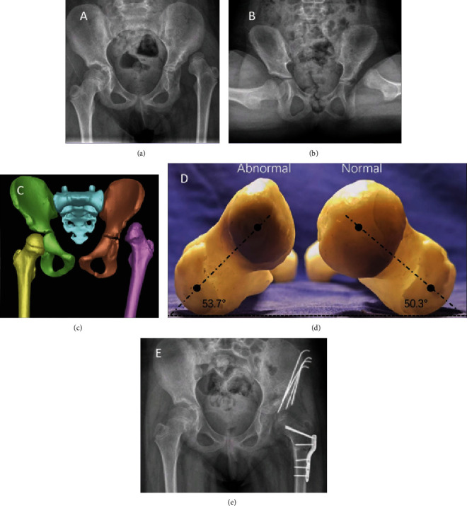
Figure 2.

The patient was a 12-year-old female with left DDH and progressive left hip pain for 1 year. She underwent left pelvic osteotomy combined with proximal femoral shortening and varus rotating osteotomy. (a, b) Preoperative radiography of the pelvis showed dislocation of the left hip joint. (c) The pelvis 3D model showed dislocation of the left hip joint. (d) Measuring the femoral neck anteversion by the femoral model. (e) Anteroposterior radiography of the pelvis after 3-month postoperation, indicating the reduction of the hip joint.
