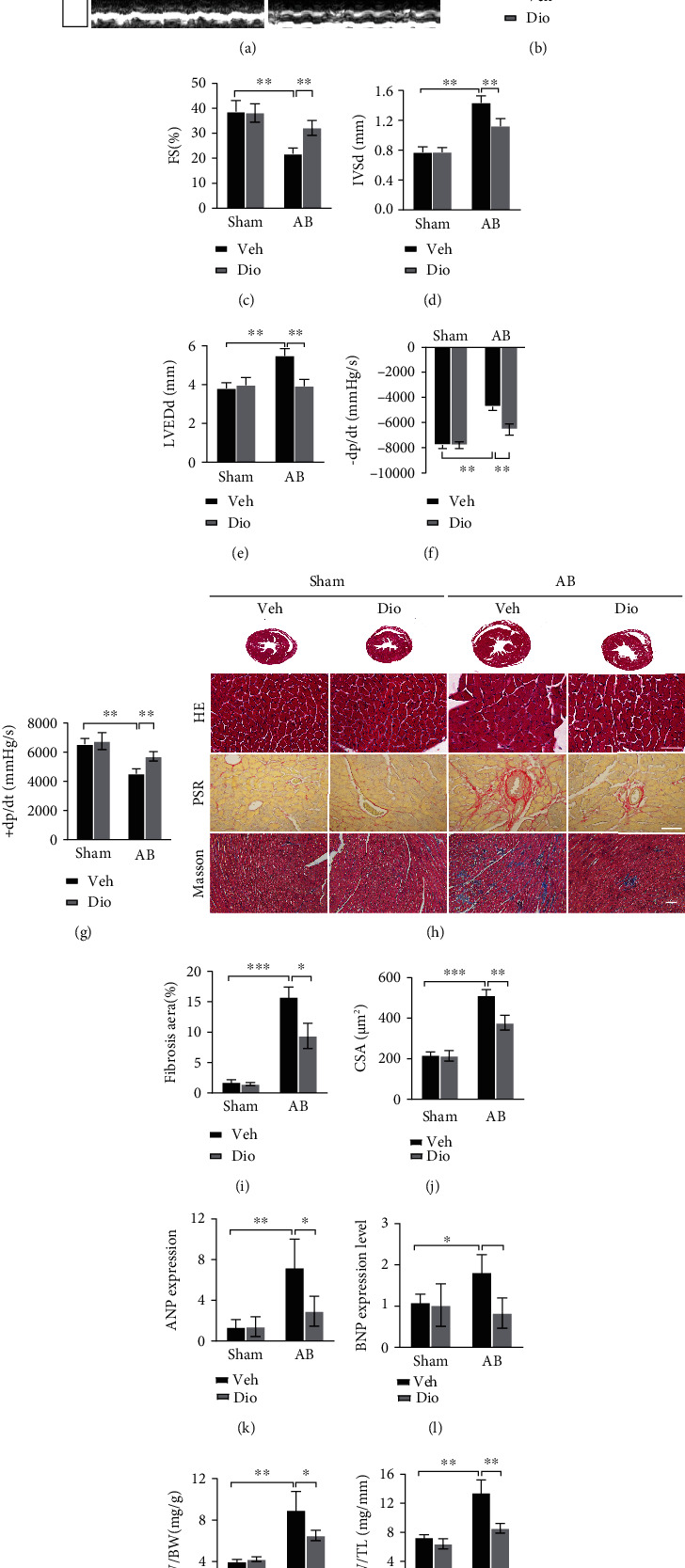
Figure 1.

Improvement of cardiac function at 4 weeks after AB by diosmetin treatment. (a) Representative M-mode images. Echocardiography was performed to measure LVEF (b), LVFS (c), IVSd (d), and LVEDd (e), n =6. (f, g) Hemodynamic parameters in suggested groups (n = 6). (h) Histopathological images of heart tissue representing Cross-sectional views of mouse hearts and the cross-sectional area of cardiomyocytes stained by HE (scale bar: 50 μm). Perivascular collagen synthesis stained by picrosirius red (PSR) (scale bar: 50 μm), and myocardial fibrosis stained by Masson trichrome staining(blue areas indicate fibrosis, scale bar: 100 μm), respectively. Quantitative of HE staining (n = 80–100) (j) and PSR staining (n = 3) (i). mRNAs for cardiac hypertrophy-associated genes ANP (k) and BNP (l) were measured by qPCR (n = 6). Gravimetric analysis of heart weight/body weight ratio (HW/BW) (m) and heart weight/tibia length (HW/TL) (n), n =6. Data are presented as mean ± SEM. ∗p <0.05, ∗∗p < 0.01, and ∗∗∗p < 0.001.
