
Figure 3.
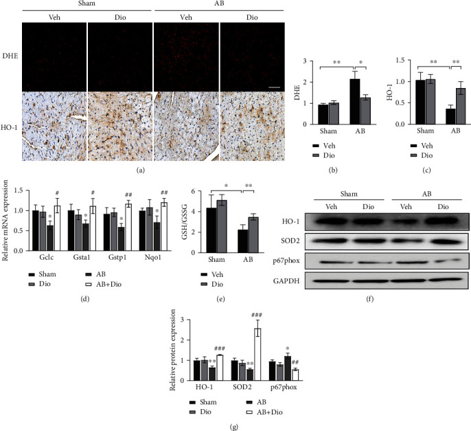
ROS reduction contributes to the protective effect of diosmetin in vivo. (a) Dihydroethidium (DHE) staining and cardiac expression of HO-1 four weeks after AB detected by immunohistochemistry, scale bar: 50 μm. Quantitative analysis of myocardial ROS levels (b) and the expression of HO-1 (c), n = 3. (d) mRNA of antioxidant enzymes Gclc, Gsta1, Gstp1, and Nqo1 mRNA in whole ventricular lysates as measured by qPCR. (e) GSH/GSSG ratios in cardiac tissues, n = 4. (f, g) Levels of HO-1, SOD2, p67phox, and GAPDH were analyzed by Western blots using GAPDH as a loading control in heart tissue lysates. Data are presented as mean ± SEM. ∗p <0.05, ∗∗p < 0.01 vs. the sham group; #p <0.05, ##p < 0.01, and ###p < 0.001 vs. the AB group; ns: not significant.