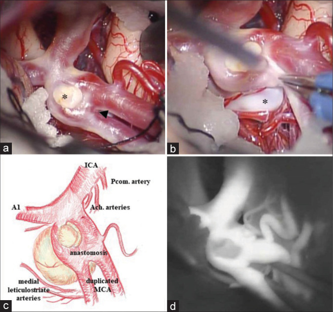
Figure 3:

The intraoperative images reveal two aneurysmal domes (asterisk) and an anastomosis (arrowhead) between d-MCA branches. The small dome protruding forward is collapsed or thrombosed (a, b: intraoperative image, c: a sketch of operative view, d: the indocyanine green video angiography.
