
Figure 2.
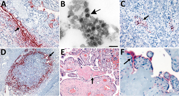
In situ hybridization (ISH) slides demonstrating localization of severe acute respiratory syndrome coronavirus 2 (SARS-CoV-2) genomic RNA in heart, liver, and lymph node tissues and electron microscopic evidence of viral particles in heart tissue from neonate in the United States that died with SARS-CoV-2 infection and placental histopathology and angiotensin-converting enzyme-2 immunohistochemical stain slides. A) SARS-CoV-2 RNA staining by nucleocapsid gene ISH assay in the endothelial cells in myocardium vessel walls (arrow). Original magnification ×20. B) Extracellular virus particles in the connective tissue of the heart (arrow). Scale bar indicates 100 nm. C) Intravascular staining by nucleocapsid gene ISH assay in the liver parenchyma (arrow). Original magnification ×20. D) Extensive nucleocapsid gene ISH staining within macrophages of subcapsular sinus of lymphoid follicle in the submucosa of upper airway (arrow). Original magnification ×10. E) Second trimester placenta with fibrinoid necrosis (arrow). Original magnification ×20. F) Angiotensin-converting enzyme 2 immunostaining in the membrane polarized on the maternal lake side in the syncytiotrophoblast (arrow). Original magnification ×63.