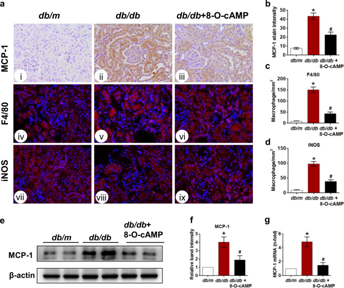
Fig. 3. Effect of the Epac agonist on MCP-1 expression and macrophage accumulation in kidney tissues.
a IHC assay assessing MCP-1 expression in the kidneys of db/m, db/db, and db/db mice treated with 8-O-cAMP (i–iii), and macrophage infiltration was visualized by F4/80 (iv–vi) and iNOS (vii–ix) immunofluorescence staining (400×). b Quantification of MCP-1 expression (n = 5). c, d Quantification of F4/80- and iNOS-positive cells per mm2 in kidney tissues (n = 5). e MCP-1 expression was evaluated by Western blotting. f Quantification of Western blot band intensities. g Relative MCP-1 mRNA expression in kidney tissues (n = 3). The values are the mean ± SD; *P < 0.05 vs db/m, #P < 0.05 vs db/db mice.

