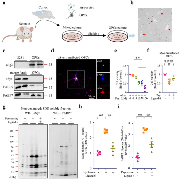
Fig. 6. FABP7 and αSyn interaction in OPCs primary culture.
a Schematic draft of OPCs primary culture protocol. b Microscopic image of OPCs (red arrows). c Western blot (WB) analysis of FABP5, FABP7, and αSyn in the U251 cells, mouse brain and in OPCs. d Confocal microscopy of immunofluorescence staining of olig2 (green), αSyn (red) and FABP7 (blue) in OPCs. e, f Cell viability analysis of OPCs, based on CCK assay. OPCs overexpressing αSyn, were treated with psychosine at various concentrations in the absence (e) (n = 6) or presence (f) (n = 8) of ligand 6. g WB analysis of FABP7 and αSyn in OPCs. OPCs, overexpressing αSyn, were treated with psychosine (10 µM) in the absence or presence of ligand 6 (1 µM). h, i Quantification of αSyn (h) and FABP7 (i) oligomers in SDS-soluble fractions, based on WB analysis, is illustrated. The data are shown as the mean ± standard error of the mean and were obtained using one-way analysis of variance. **P < 0.01 and ##P < 0.01. ɑSyn alpha-synuclein, FABP3, 5, and 7 fatty acid-binding protein 3, 5, and 7, Psy Psychosine.

