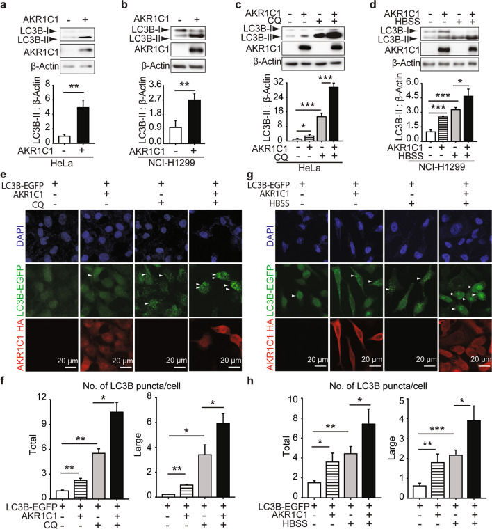
Fig. 4. AKR1C1 accelerates autophagy flux.
HeLa (a and c) or NCI-H1299 cells (b and d) were transfected with AKR1C1 or vector plasmid and assessed by Western blotting. Band intensities of LC3B-II were measured and normalised to β-Actin. c HeLa cells were exposed to CQ (10 μM) for 15 h at 40% confluency. d NCI-H1299 cells were cultured in HBSS for 2 h. e–h LC3B-EGFP stably-expressed HeLa cells were constructed and cultured for 5 generations. These cells were transfected with AKR1C1 and treated by CQ (10 μM, 15 h) or HBSS (2 h). Then cells were subjected to IF. LC3B puncta were quantified by Image J. Green: LC3B-EGFP. Red: AKR1C1. Blue: cell nuclei. Scale bar: 20 μm. Large: size ≥ 5.0 pixel units. Total: size ≥ 2.5 pixel units.

