Fig. 4.
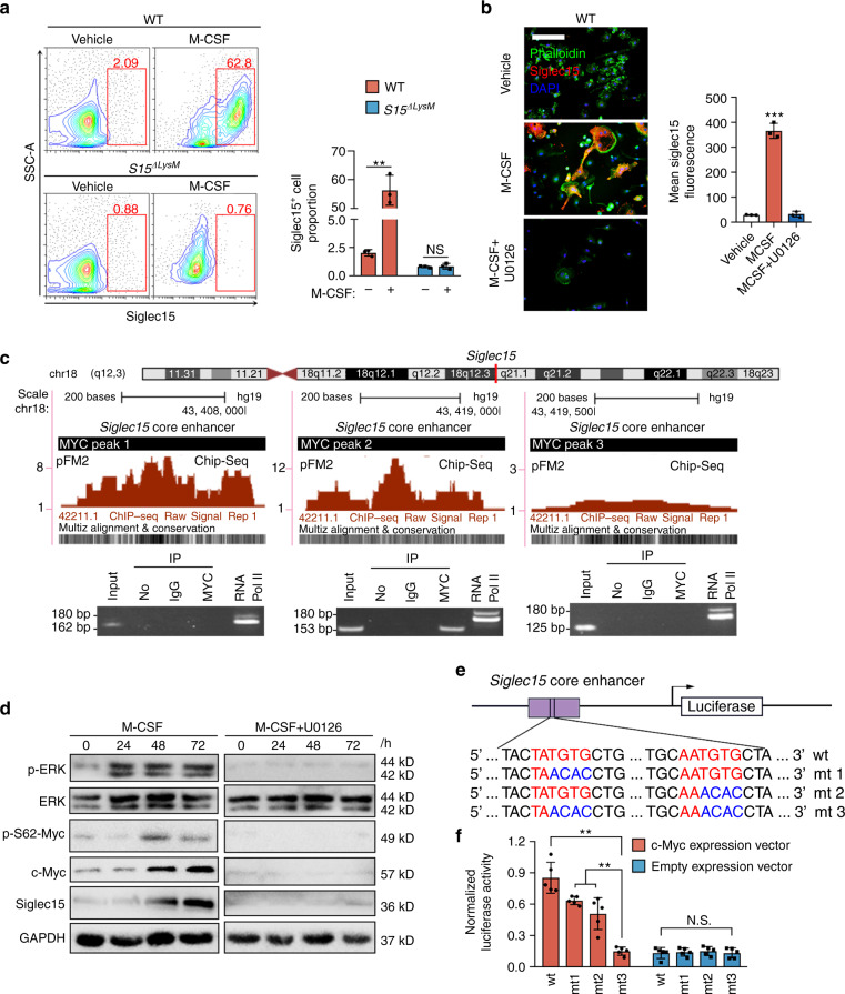
Siglec15 expression in macrophages/monocytes is induced by M-CSF via MEK-ERK-MYC signaling. a Flow cytometric analysis of Siglec15fl/fl (WT) and Siglec15ΔLysM BMMs treated with M-CSF or vehicle for 3 days, with quantification of the Siglec15+ cell proportion. b Immunostaining of BMMs treated with M-CSF, M-CSF + U0126 or vehicle with phalloidin and anti-Siglec15, with quantification of Siglec15+ cells; the bar represents 50 μm. c Three MYC binding sites in the Siglec15 promoter core enhancer region and chromatin immunoprecipitation (ChIP) assay validation of the MYC binding sites. d Western blot analysis of ERK, MYC phosphorylation, and Siglec15 in BMMs treated with M-CSF, M-CSF + U0126 or vehicle at 0, 24 h, 48 h, and 72 h. e Site-directed mutagenesis of the MYC binding sites in the Siglec15 core enhancer. f Quantification of the normalized luciferase activity of a luciferase gene reporter assay performed with BMMs transfected with a c-Myc expression vector, n = 5. Data represent the mean ± SD, and statistically significant differences are indicated as **P < 0.01; ***P < 0.001

