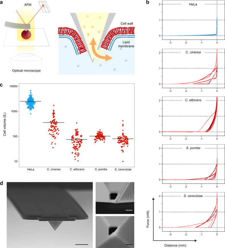
Fig. 1. Technological developments.
a Scheme of FluidFM-based injection/extraction of fungi. A microfluidic probe operated with an atomic force microscope (AFM) is inserted through the hard cell wall, allowing for a pressure-driven exchange of liquids between the probe microchannel and the cell interior. The FluidFM is mounted on an optical microscope that allows in situ monitoring of the manipulation. b Representative force-distance curves obtained upon probe insertion in the different organisms. Each plot features 5 curves recorded with different cells. c Estimated cell volumes, as measured from 2D microscopy images assuming tubular and spherical cell geometries, for (dissociated) HeLa cells (N = 132), C. cinerea (N = 88), C. albicans hyphal compartments (N = 116), S. pombe (N = 100) and S. cerevisiae (N = 105). Horizontal lines are means. d Scanning electron microscopy images of FluidFM probes with a custom-designed tip aperture for fungal injection. Left: Front view of the hollow cantilever with a pyramidal tip. Scale bar: 5 µm. Right: side- (top) and bottom- (down) views of the aperture on the pyramidal tip. Scale bars: 200 nm.

